
NeXT - Neutron and X-ray Tomography instrument

NeXT (Neutron and X-ray Tomography) is a cold neutron imaging instrument with additional X-rays imaging capabilities. These two modalities can be operated simultaneously.
The typical configurations allow neutron radiography and tomography with fields of views ranging from 4.1×4.1 mm2 to 170×170 mm2.
NeXT is located at the end of the H521 guide shared with MoTo, D16 and SuperADAM.
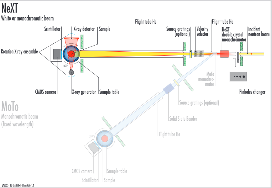
Instrument layout
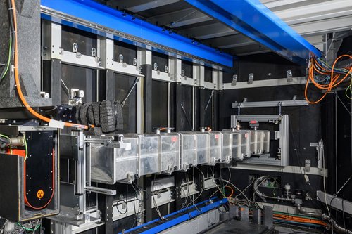
The neutron beam passes through a set of adjustable pinhole of diameter D that acts as a primary source. The diverging beam travels a distance L of 4.5-11 m to arrive to the Imaging station, where the sample is mounted. The pinhole selection and movable sample station allow one to change beam intensity and collimation ratio L/D.
The imaging station is equipped with a rotation table to allow for tomographic imaging (including laminography). Behind the sample, a set of possible detectors acquire transmission images of the sample.
Perpendicular to the neutron beam, an ensemble X-ray generator / detector allows the acquisition of complementary X-ray tomographies of the same sample.
Wavelength selective imaging is available by means of a double crystal monochromator as well as a velocity selector. Additional equipment can be installed along the beam to allow for example polarised neutron imaging, grating interferometry, scattering correction mask.
Techniques available:
- White-beam neutron imaging at spatial resolutions down to 4 μm
- Wavelength selective imaging:
- Double crystal monochromator (2-6 Å)
- Velocity selector (2-20 Å)
- Bragg-edge imaging
- Polarised neutron imaging
- Grating interferometry
- Laminography
- Complementary X-ray imaging (20 to 300 kV source), with spatial resolutions down to approx. 4 microns
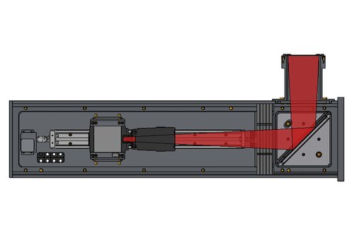
Imaging detector - the detector aimed to have maximum flexibility of the field of view and corresponding resolution. The light emitted by the scintillator is reflected vertically by a mirror inclined by 45º. The light is then captured by a sCMOS camera (2048x2048 pixels) mounting a variety of lenses, and which moves on a rail. This allows for modifying the field of view, which ranges from 170x170 mm2 at a resolution of about 100µm to a field of view of 20x20 mm2 and a resolution of about 30 µm. A second detector is currently under development, which allows for higher resolutions, down to 10 µm. Accordingly a set of scintillators is employed, ranging from a 200 µm thick ZnS/Li6F scintillator to a 2.5 µm thick 157Gd2O2S:Tb scintillator.
Applications
- Energy storage like Li-ion batteries and fuel cells
- Porous media
- Earth sciences
- Materials Science
- Engineering and metallurgy
- Cultural and Natural Heritage
- Water transport in plants or porous media
- Wavelengths selective imaging to exploit diffraction contrast (Bragg edges) of different materials
- Magnetic fields and domains
NeXT is supported by the Auvergne Rhône-Alpes (AURA) region
