D33 - Massive dynamic q-range small-angle diffractometer
D33 is a Small-Angle Neutron Scattering instrument for the characterization of samples with typical sizes varying from the nanometer scale to few tenth of micrometer. In addition to a standard monochromatic mode of operation, D33 offers a time of flight mode (TOF) to cover an enhanced dynamic q-range qmax/qmin in one instrument setting.
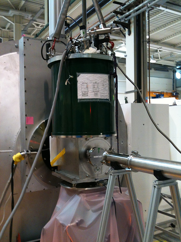
High magnetic fields, up to 17 T at the sample position, beam polarization and 3He spin analysis, facilitate and expand studies of magnetism and allow a more quantitative analysis of spin incoherent samples. The high flux allows for kinetic experiments with time resolution of the order of few milliseconds.
For materials science and physics studies, the usual array of sample environments, e.g. electromagnet, cryostat, cryomagnet and furnace, can be mounted easily on D33. The generous sample space can accommodate bulky and massive sample environments such as large cryomagnets, e.g. the 17 T cryomagnet belonging to the University of Birmingham, as well as 3He spin analysis setup.
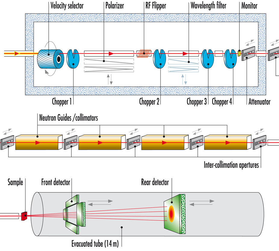
In the scheme, the instrument layout is shown in 3 parts:
- Top: neutron selection either with a neutron velocity selector (monochromatic mode) or with choppers (time of flight mode)
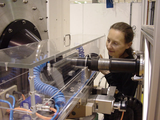
- Middle: collimation part
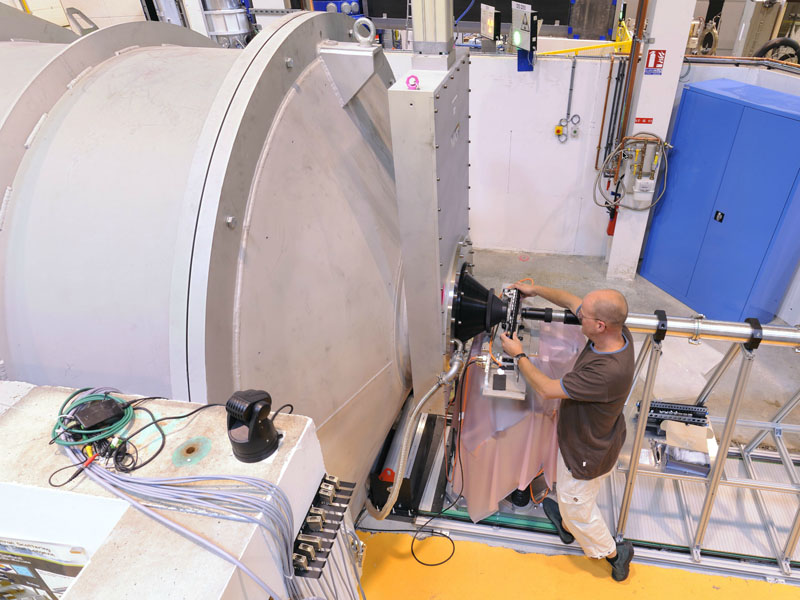
- Bottom: front and rear detector moving in the vacuum tube from 1.2 m to 12.8 m after the sample position.
Applications
- Soft condensed Matter:
organic and inorganic colloidal particles; polymers blends or polymer in solution, gels, liquid crystals; self assembly of surfactant molecules. - Biology:
proteins, nucleic acids, biomembranes, vectors for drug delivery. - Material Science:
phase separation in alloys and glasses, morphologies of superalloys, microporosity in ceramics, interfaces and surfaces of catalysts. - Magnetism:
Flux line lattices in superconductors; magnetic correlations