More
Miscellaneous
D19 - Introduction
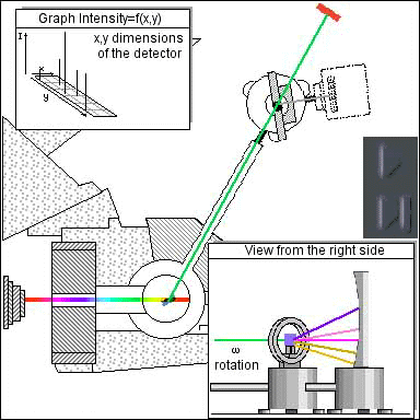
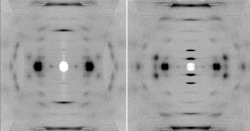
D19 is a diffractometer combining a high flux monochromatic neutron beam and a large area detector that covers Horizontal 120° x Vertical 30°. This provides the user community with the only instrument capable of surveying reciprocal space for small samples with large unit cells, for d-spacings from 100Å to 0.5Å. The instrument has proven applications in biology, chemistry, physics, materials science and polymer science. It is the only instrument at the ILL that can record single crystal diffraction data to atomic resolution from small samples with relatively large unit cells; it is also the only instrument in the world that can perform high-angle neutron fibre diffraction experiments.
The instrument has been used to carry out single crystal studies of systems such as vitamin B12, lysozyme, haemoglobin. In fibre diffraction mode, D19 has been used to investigate hydration structure around various conformations of DNA, hyaluronic acid, cellulose, and filamentous viruses. It has also been used to investigate the conformation of industrial polymer systems, including high-performance thermoplastics.
D19 for single crystal science
In single crystal work on small molecule systems, D19 allows the determination of the location of light atoms amongst heavy ones and the accurate characterisation of liganded H and H2 and of hydrogen bonding and hydrogen disorder in studies of organic and inorganic molecules, complexes, solvates, and adducts.
Some larger molecules of biological interest have also been studied on D19, such as vitamin B12 (Jogl et al., in preparation), lysozyme (Bouquiere et al., 1993b), and haemoglobin (Waller, 1989). The interest here is obvious – information on proton positions and hydration is vital for understanding biological processes. Data from monochromatic neutron diffraction studies allows H or D positions to be determined at a much higher level of significance than is obtainable with x-rays. Although it is true that such information can be obtained from high resolution x-ray diffraction when crystals diffract to 1.2Å or better, it is also true that only a small fraction of crystals diffract to this resolution.
Crystallography
- Incommensurate structures of the [CH3NH3][Co(COOH)3] compound, Cañadillas-Delgado L., Mazzuca L., Fabelo O., et al ; IUCrJ 6, 105-115 (2019) doi:10.1107/S2052252518015026
- Hydrogen-bonding network in anhydrous chitosan from neutron crystallography and periodic density functional theory calculations, Ogawa Y., Naito P.K., Nishiyama Y. ; Carbohydrate Polymers 207, 211-217 (2019) doi:10.1016/j.carbpol.2018.11.042
- Microscopic insights on the multiferroic perovskite-like [CH3NH3][Co(COOH)3] compound, Mazzuca L., Cañadillas-Delgado L., Fabelo O., et al ; Chemistry - A European Journal 24, 388-399 (2018) doi:10.1002/chem.201703140
- London dispersion enables the shortest intermolecular hydrocarbon H···H contact, Rosel S., Quanz H., Logemann C., et al ; Journal of the American Chemical Society 139, 7428-7431 (2017) doi:10.1021/jacs.7b01879
- High resolution X-ray and neutron diffraction studies on molecular complexes of chloranilic acid and lutidines, Sovago I., Thomas L.H., Adam M.S., et al ; CrystEngComm 18, 5697-5709 (2016) doi:10.1039/C6CE01065B
- Nature of Si-H interactions in a series of ruthenium silazane complexes using multinuclear solid-state NMR and neutron diffraction, Smart K.A., Grellier M., Coppel Y., et al ; Inorganic Chemistry 53, 1156-1165 (2014) doi:10.1021/ic4027199
- CNN pincer ruthenium catalysts for hydrogenation and transfer hydrogenation of ketones: Experimental and computational studies, Baratta W., Baldino S., Calhorda M.J., et al ; Chemistry - A European Journal 20, 13603-13617 (2014) doi:10.1002/chem.201402229
- Host perturbation in a β-hydroquinone clathrate studied by combined X-ray/neutron charge-density analysis: Implications for molecular inclusion in supramolecular entities , Clausen H.F., Jørgensen M.R.V., Cenedese S., et al ; Chemistry - A European Journal 20, 8089-8098 (2014) doi:10.1002/chem.201400129
- Neutron diffraction characterization of C-H···Li interactions in a lithium aluminate polymer , Cole J.M., Waddell P.G., Wheatley A.E.H., et al ; Organometallics 33, 3919-3923 (2014) doi:10.1021/om500271p
- SPINE-compatible 'carboloops': A new microshaped vitreous carbon sample mount for X-ray and neutron crystallography, Romoli F., Mossou E., Cuypers M., et al ; Acta Crystallographica F 70, 681-684 (2014) doi:10.1107/S2053230X14005901
Magnetism
- Direct observation of finite size effects in chains of antiferromagnetically coupled spins, Guidi T., Gillon B., Mason S.A., et al ; Nature Communications 6, 7061-1-7061-6 (2015) doi:10.1038/ncomms8061
Geoscience, Materials Science and Engineering
- Single-crystal neutron diffraction study of wardite, NaAl3(PO4)2(OH)4·2H2O, Gatta G.D., Guastoni A., Fabelo O., et al ; Physics and Chemistry of Minerals 46, 427-435 (2019) doi:10.1007/s00269-018-1013-7
- Volume texture and anisotropic thermoelectric properties in Ca3Co4O9 bulk materials, Kenfaui D., Chateigner D., Gomina M., et al ; Materials Today: Proceedings 2, 637-646 (2015) doi:10.1016/j.matpr.2015.05.089
- Submarine lava flow direction revealed by neutron diffraction analysis in mineral lattice orientation, Zucali M., Fontana E., Panseri M., et al ; Geochemistry, Geophysics, Geosystems 15, 765-780 (2014) doi:10.1002/2013GC005044
- Zucali M., Barberini V., Voltolini M., et al ; Quantitative 3D microstructural analysis of naturally deformed amphibolite from the Southern Alps (Italy): Microstructures, CPO and seismic anisotropy from a fossil extensional margin Geological Society, London, Special Publications 409, 201-222 (2014) doi:10.1144/SP409.5
D19 for Biology, Fiber and Soft Matter
The availability of an area detector has also meant that D19 can be used to record high-angle neutron fibre diffraction patterns. The first experiments of this type were on DNA hydration and provided the first information on hydration in polymeric DNA. The studies are also important because fibres allow the study of DNA conformations that have not been observed in oligonucleotide single crystals and also of stereochemical changes that occur during conformational transitions (see Shotton et al., 1997).
The same techniques have since been used to study hyaluronic acid, filamentous viruses, and have recently produced some outstanding results from cellulose (Nishiyama et al., 1999; Langan et al., 1999). Similar methods have been used to study hydrogen atoms in aromatic polymers (Mahendrasingam et al., 1992) and are currently being developed for the study of polymers such as Nylon 66. There is increasing interest from industrial collaborators in the use of fibre diffraction methods in combination with specific deuteration to study changes in polymer structure as a function of chemical composition, temperature, and drawing processes. All of the neutron fibre diffraction studies provide information that cannot be obtained from complementary x-ray fibre diffraction studies.
Biology
- Inner-sphere water and hydrogen bonds in lanthanide DOTAM complexes. A neutron diffraction study, Bombieri G., Artali R., Mason S.A., et al ; Inorganica Chimica Acta 470, 433-438 (2018) doi:10.1016/j.ica.2017.09.021
- Macromolecular structure phasing by neutron anomalous diffraction , Cuypers M.G., Mason S.A., Mossou E., et al ; Scientific Reports 6, 31487-1-31487-7 (2016) doi:10.1038/srep31487
- Biomolecular deuteration for neutron structural biology and dynamics, Haertlein M., Moulin M., Devos J.M., et al ; Methods in Enzymology 566, 113-157 (2016) doi:10.1016/bs.mie.2015.11.001
- Neutron diffraction studies on guest-induced distortions in urea inclusion compounds, Lee R., Mason S.A., Mossou E., et al ; Crystal Growth & Design 16, 7175-7185 (2016) doi:10.1021/acs.cgd.6b01371
- Looking for hydrogen atoms: Neutron crystallography provides novel insights into protein structure and function, Golden E.A., Vrielink A.; Australian Journal of Chemistry 67, 1751-1762 (2014) doi:10.1071/CH14337
- Binding site asymmetry in human transthyretin: Insights from a joint neutron and X-ray crystallographic analysis using perdeuterated protein, Haupt M., Blakeley M.P., Fisher S.J., et al ; IUCrJ 1, 429-438 (2014) doi:10.1107/S2052252514021113
- L-arabinose binding, isomerization, and epimerization by D-xylose isomerase: X-ray/neutron crystallographic and molecular simulation study, Langan P., Sangha A.K., Wymore T., et al ; Structure 22, 1287-1300 (2014) doi:10.1016/j.str.2014.07.002
- Structure and spacing of cellulose microfibrils in woody cell walls of dicots, Thomas L.H., Forsyth V.T., Martel A., et al ; Cellulose 21, 3887-3895 (2014) doi:10.1007/s10570-014-0431-z
- SPINE-compatible 'carboloops': A new microshaped vitreous carbon sample mount for X-ray and neutron crystallography, Romoli F., Mossou E., Cuypers M., et al ; Acta Crystallographica F 70, 681-684 (2014) doi:10.1107/S2053230X14005901
Soft Matter
- Melting transition of oriented DNA fibers submerged in poly(ethylene glycol) solutions studied by neutron scattering and calorimetry, González A., Wildes A., Marty-Roda M., et al ; Journal of Physical Chemistry B 122, 2504-2515 (2018) doi:10.1021/acs.jpcb.7b11608
- Diffraction evidence for the structure of cellulose microfibrils in bamboo, a model for grass and cereal celluloses, Thomas L.H., Forsyth V.T., Martel A., et al ; BMC Plant Biology 15, 153-1-153-7 (2015) doi:10.1186/s12870-015-0538-x
- Solid-solvent molecular interactions observed in crystal structures of β-chitin complexes, Sawada D., Ogawa Y., Kimura S., et al ; Cellulose 21, 1007-1014 (2014) doi:10.1007/s10570-013-0077-2
Considering an experiment?
Instrument D19 is a neutron 4-circle diffractometer that can be used to carry out high resolution work on relatively small single crystals that have large unit cells, and is routinely used to carry out such work of interest in biological systems, materials science, physics, and chemistry.
The instrument has a multiwire area detector. This also allows high-angle neutron fibre diffraction experiments to be carried out and a significant fraction of beamtime on D19 is awarded for experiments on biological and synthetic polymer systems including nucleic acids, filamentous bacteriophages, cellulose, hyaluronic acid, and industrial polymers.
If you are considering an experiment on D19, please do not hesitate to contact either instrument responsible :
Estelle Mossou (mossou(at)ill.fr, or phone +33 (0) 4 76 20 94 41)
Laura Canadillas Delgado (canadillas-delgado(at)ill.fr, or phone +33 (0) 4 76 20 79 34)
or for technical questions (sample mounting etc), you can contact the instrument technician for D19 and D9 :
John Archer (archer(at)ill.fr or phone +33 (0) 4 76 20 74 22)
who will be more than happy to discuss the work that you are interested in doing and the best way forward.
Other ILL scientists with detailed knowledge of D19 are :
Trevor Forsyth (tforsyth(at)ill.fr)
Oscar FabeloRosa (fabelo(at)ill.fr)
Bachir Ouladdiaf (ouladdiaf@ill.fr)
There are a number of different diffractometers at the ILL and if D19 is not the best one for your work, we will point you in the right direction. Check this link for information on the other single crystal diffraction instruments:
Diffraction group single crystal diffractometers (D3, D9, D10, D23)
Longer wavelength diffractometers (in the Large Scale Structures group) (D16, LADI)
See also general information on the ILL, and specfic information on how to submit a proposal.
Before coming to the ILL
Before coming to the ILL
You (and we) will not get the most out of your experiment if you are not properly prepared. You should consider the following issues carefully before you arrive. Talk to your local contact if you have any doubts.
Sample
- If the crystal volume is smaller than in your proposal or less than 1 mm3 per 1000A3 - tell your local contact at least 4 weeks before the experiment, and consider whether to postpone or cancel the experiment.
- Choose the compound and solvent carefully. Avoid if possible (mixed) organic solvents - high H content - which may give disorder complicating or preventing refinemnt
- Check the effect of cooling (or heating): phase transitions, cracking,
- Select at least 3 suitable crystals (or fibres...): quality,size, shape...
- How to mount a crystal NYA
- Determine the dimensions of the 3 best crystals: perpendicular distance from an arbitrary internal origin to each plane face. If this is not done, attempt on Huber optical goniometer at ILL before start of experiment.
- Index the faces e.g. on smart in home lab - this will avoid wasting neutron time doing it on d19 at start of the experiment.
Background information
- Refine the x-ray structure carefully - at the temperaure of the neutron expt if possible - anisotropic TF's, solvent, non-controversial H atoms (yes !)
- Calculate neutron F**2 for hkl's up to e.g. 2-theta=80 at e.g. 1.31A. You can even sort them on F**2 with Unix sort command: -e.g. sort -r +3 -4 unsorted.fcf -osorted.fcf
- Where unsorted.fcf is the fcf file from shelxl with header stuff removed
- E-mail F**2 list and e.g. *.ins or xtal input file to your local contact
- Check the ILL web page at wwwold.ill.fr, for info on telnet connection, etc
- Learn a few basic Unix commands; we use an editor such as nedit (or vi ugh!)
D19 computers (in progress)
There are a number of computers that are available to you as a visitor to instrument D19. Two of these are located in the instrument area - you have to be familiar with their operation in order to execute you experiment and evaluate your data. Their details are summarised below :
Computer name | Network name | Function |
|---|---|---|
d19 | d19.ill.fr | Data acquisition using NOMAD |
d19lnx | d19lnx.ill.fr | Data reduction & analysis |
As soon as you arrive, you should set yourself up on each of these computers as follows:
Log in as d19. You will then be forced to work in a sub-directory - please use your user-club identifier
example
Enter your surname : chadwick
you are now in /home/vis/d19/chadwick
d19
Run NOMAD in this area.
Don't work in the main area /users/d19 unless necessary.
User log files are available onlogs.ill.fr. You can enrich them with comments, pictures, files...
d19lnx
Run all data reduction & analysis software from here (e.g. for single crystal experiments, programs such as pfind, retreat, rafd19, d19abs, d19abscan etc).
N.B.: - Remember that linux differentiates between lower and upper case: best never to use capital letters unless required
- use ctrl-c not ctrl-z to stop most programs - this minimises detached jobs. However, do not use ctrl-c to stop NOMAD - it may crash the computer - use pcp.
Data Reduction & Analysis Softwares (in progress)
Instrument control
Program | Function |
|---|---|
| nomad | from 2019, Instrument control program |
| mad | until 2018, Instrument control program (uses the prompt 'MT |
Data reduction
| Program | Function |
|---|---|
| pfind | search peaks in a set of scans/numors |
| index | Index reflections that have been found |
| rafd19 | Calculate UB matrix, cell dimensions, offsets etc |
| retreat | integration of peaks after indexing |
| compact | Checking retreat output |
| d19abs | Calculate absorption correction for each hkl |
| d19abscan | Apply absorption and "can" correction |
Some useful Unix commands
| Command | Effect |
|---|---|
| bg | Put a foreground job into the background |
| fg | Bring a background job into the foreground |
| find /users/dif/mason/progs -name "hklcol" | Print to locate hklcol |
| find /tmp_mnt/home/vis/d19 -name "*.f" | Print to locate *.f |
| find ../ -name "hklcol" | Print to locate prog hklcol starting from directory above present |
| grep "comhkl" ~mcintyre/progs/peakint/std/* | Find string "comhkl" in history list of commands used in present session |
| gedit filename | Text editor on Silicon Graphics machines and on DEC alpha |
| history | List of ~100 last commands |
| kill -9 1234 | Kill process no. 1234 |
| lp filename | Print on default d19sgi printer at instrument (i.e. hp_d19) |
| ls /hosts/d19/users/data | List files in directory |
| man program | Help on the program |
| ps | Find out which processes are running |
| ps -edf |grep models | Find out more about process e.g. models |
| q | Exit from man, more, etc |
| retreat > retreat.log & | Will run retreat in background, so can logout. Be sure to first do setenv DI1 .... etc |
| setenv DI1 /hosts/d19/users/data | Force retreat to take data fr d19 or setd19 (or setserdon to take transferred data from serdon) |
| setenv DI1 /hosts/serdon/illdata/data-n/974/d19 | Force retreat to find old cycle 974 data (first do ls to check available -see ls) |
| source ../.cshrc.group | Force execution of that file (if not automatic) |
| which anything | Find out what anything is defined as (aliases, progs..) |
Instrument construction
EPSRC project for chemical crystallography and fibre diffraction using the ILL diffractometer D19


An award from the UK Engineering and Physical Sciences Research Council (EPSRC) has provided resources for a new large area position-sensitive detector on the D19 diffractometer at the Institut Laue Langevin (ILL). The modified instrument will have a detection solid angle that is larger by a factor of ~25 than in the current configuration. In addition to providing vast enhancements in quality for experiments of the type that are currently carried out, the upgraded instrument will allow completely new science to be undertaken.
This development is of broad interest for neutron crystallography and fibre diffraction and for applications of these methods in chemistry, biology and physics. These communities have identified a clear need for a flexible monochromatic instrument that can carry out rapid neutron diffraction analyses to high resolution. The large increase in detection solid angle means that it will be possible to study the small single crystal samples most frequently encountered in this field and by exploiting tighter incident beam coIlimation, to investigate larger unit cells. For fibre diffraction work there will be the added advantage of being able to measure continuous diffraction.
The new detector
The new detector is being built by the ILL Detector Group and will be commissioned in mid 2005. The drawings shown below summarise the design and show how the detector will be installed.


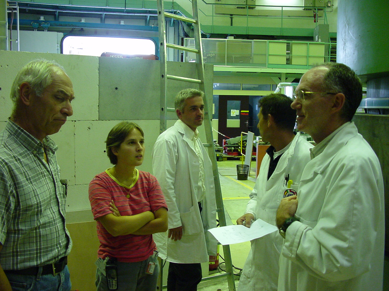
Below: the complete detector (all except the electronics) just before being sealed in its vacuum vessel. Seen with it are Jean-Francois Clergeau, Sax Mason, Clive Wilkinson, Arsen Goukassov.

Applications
Chemistry & Materials
- Charge density analysis
- Accurate structure determination for crystal engineering
- Studies of transition metal catalysis
- Structural studies of new organic materials
- Characterisation of weak intermolecular interactions involving hydrogen
- Structure-property studies of opto-electronic/magnetic materials
- Synthetic polymers
Biology
Biopolymers :
- nucleic acids
- filamentous viruses
- cellulose
- chitin
- amyloid fibres
Biological crystallography :
- Small proteins
- Oligonucleotides
Examples
Revised structure for Kevlar, based on neutron fibre diffraction analysis ( Gardner, English, Forsyth, Macromolecules 37, 9654-9656 (2004) (pdf - 332 Ki))
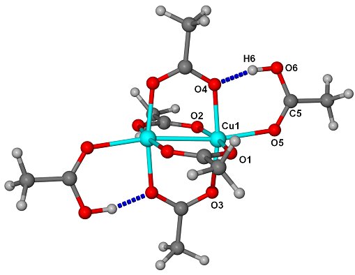
Structures of [Cu2(m-O2CMe)4(O2HCMe)2] determined as part of an investigation of the effect of strong intra- and inter- molecular hydrogen bonding on molecular geometry in carboxylate coordination complexes (Vives et al (2001).

References
Ahrens B, Davidson MG, Forsyth VT, Mahon MF, Johnson AL, Mason SA, Price RD, Raithby PR, J. Am. Chem. Soc., 9164-9165, 123, (2001)
Broder CK, Davidson MG, Forsyth VT, Howard JAK, Lamb S, Mason SA, Cryst. Growth Des., 163-169, 2, (2002)
Gardner,K.H., English, A.D., Forsyth, V.T., Macromolecules 37, 9654-9656 (2004) (pdf - 332 Ki)
Langan, P. , et al, J. Am. Chem Soc. 121 (43) 9940-9946 (1999).
Nishiyama, Y. , et al, Int. J. Biol. Macr. 26 (4), 279-283 (1999).
Vives,G., Mason, S.A., Prince, P.D., Junk, P. Steed, J.W., Crystal Growth & Design, 3, 699 (2003)
Ressources
Estimating activation
Using the following procedure and example, estimate the anticipated activation of a sample exposed to the neutron beam in a LANSCE instrument from the table. If you cannot estimate the activation, call your LANSCE contact.
Example: For 5g YBa2Cu3O7 sample on NPD at 75mA for 24 hours (1 day).
1. Compute the mass fractions for all elements in the sample.
Example: The mass fractions are 0.13 Y, 0.4l Ba, 0.29 Cu, & 0.17 O
2. Obtain the prompt activation from the product of these mass fractions and the corresponding elemental activation values in the table. Multiply the contact dose by the sample mass that is exposed to the beam to obtain the expected contact dose from the entire sample.
Example continued:
prompt activity = 0.13x 1000 + 0.41x80 +0.29x 10000 + 0.17x0.0 = 3060nCi/g
contact dose = 5x(0.13x0.9 + 0.41x0.1 + 0.29x8.5 + 0.17x0.0) = 13.1mR/hr
3. Scale these activation values by the instrumental factor (below) and the beam current as a fraction of l00mA to obtain the estimated values for the sample and actual beam conditions.
Instrumental factors for LANSCE
HIPD 1.00 (reference)
SCD 1.44
FDS 0.50
CQS 1.65
NPD 0.025
PHAROS 0.060
LQD 0.033
SPEAR 0.04 1
Example continued:
prompt activity = 3060x0.025x75/100= 57nCi/g
contact dose = 13.lx0.025x75/100 = 0.25mR/hr
4. To obtain the storage times, t, for each element in the sample, correct the appropriate times given in the table, ts, by:
t = ts(0.693+0.lxlogeC),
where C is the product of the mass fraction, instrumental factor and beam intensity ratio to l00m A. This expression assumes that the storage times given in the table are roughly 10 half-lives for the slowest decaying isotope of significant concentration in the activated element The storage time for the sample is then the largest of the resulting times.
Example continued
Y storage time = 24d(0.693+0.lxloge(0.13x0.025x75/100)) = 2.2d
Ba storage time = 150h(0.693+0.lxloge(0.41x0.025x75/100)) = 1.3d or less
Cu storage time = 7.4d(0.693+0.lxloge(0.29x0.025x75/100)) = 1.3d
O storage time = 0 (no activation expected)
Thus the storage time for this sample of YBa2Cu3O7 is determined by the decay of the Y as 2.2 days. At that time, the sample should show residual activation of less than 2nCi/g.
These estimates are based on a neutron exposure time of 1 day. The actual level of activation can drastically change if the exposure is more or less than 1 day. Determining this change cannot be accomplished using these simplistic calculations. Therefore, we require that all samples (no matter how long the neutron exposure is) be surveyed before they leave the facility.
LANSCE 12 / 1 / 92
Activation table of the elements
This table allows you to calculate the activation of a sample after it has been in a neutron beam for one day and the amount of time for it to decay to 2nCi/g or less, which is the limit for shipping a sample as "nonradioactive". It also displays the anticipated exposure you may receive when removing the sample from the instrument. A sample calculation is included at the end of the table. Storage time is the time required for a sample of the pure condensed-phase element exposed to a "standard" neutron beam to decay to 2nCi/g or less. Prompt activation gives the anticipated activation for the pure solid elements 2 min after the neutron exposure ceases. Contact dose is that expected from a 1g sample of the pure element from the prompt activation. Elements with a dash for the entries in all three columns do not show any activation. Those marked with a single asterisk are radioactive before exposure to the neutron beam; apart from Tc and Pm, they are all a-particle emitters. Bismuth is a special case; it is stable before exposure to the beam, but the activation product is an a-emitter.
| Symbol | Name | Mass | Storage time | Prompt activation | Contact dose |
|---|---|---|---|---|---|
| (nCi/g) | (mr/hr/g at 1 in) | ||||
| Ac | actinium | 227 | * | * | * |
| Al | aluminium | 26.982 | 21m | 1900 | 2.0 |
| Am | americium | 243 | * | * | * |
| Sb | antimony | 121.75 | 520d | 800 | 0.7 |
| Ar | argon | 39.948 | 19h | 3500 | 3.0 |
| As | arsenic | 74.922 | 18d | 8.4x10⁴ | 7.3 |
| At | astatine | 210 | * | * | * |
| Ba | barium | 137.34 | <150h | <80 | <0.1 |
| Bk | berkelium | 247 | * | * | * |
| Be | beryllium | 9.012 | - | - | - |
| Bi | bismuth | 208.980 | ** | ** | ** |
| B | boron | 10.811 | - | - | - |
| Br | bromine | 79.909 | 18d | 1.4x10⁴ | 12† |
| Cd | cadmium | 112.40 | 190d | 370 | 0.3 |
| Ca | calcium | 40.08 | - | - | - |
| Cf | californium | 249 | * | * | * |
| C | carbon | 12.011 | - | - | - |
| Ce | cerium | 140.12 | <86h | <40 | <0.1 |
| Cs | cesium | 132.905 | 54h | 4.6x10⁵ | 400 |
| Cl | ch1orine | 35.453 | <2.8h | <80 | <0.1 |
| Cr | chromium | 5 1.996 | <6ld | <40 | <0.1 |
| Co | cobalt | 58.933 | 24y | 5.2x10⁴ | 45† |
| Cu | copper | 63.54 | 7.4d | 1.0x10⁴ | 8.5 |
| Cm | curium | 247 | * | * | * |
| Dy | dysprosium | 162.50 | 52h | 5.0x10⁵ | 430† |
| D | deuterium | 2.015 | - | - | - |
| Es | einsteinium | 254 | * | * | * |
| Er | erbium | 167.26 | 78d | 600 | 0.5 |
| Eu | europium | 151.96 | 50y | 2200 | l.9† |
| Fm | fermium | 253 | * | * | * |
| F | fluorine | 18.998 | - | - | - |
| Fr | francium | 223 | * | * | * |
| Gd | gadolinium | 157.25 | 11d | 7400 | 6.4 |
| Ga | gallium | 69.72 | 8d | 3.2x10⁴ | 27† |
| Ge | germanium | 72.59 | <6d | 1100 | 1.0† |
| Au | gold | 196.967 | 29d | 3000 | 2.5 |
| Hf | hafnium | 178.49 | 1.6y | 620 | 0.5 |
| He | helium | 4.003 | - | - | - |
| Ho | holmium | 164.930 | 20d | 2.8x10⁴ | 24† |
| H | hydrogen | 1.008 | - | - | - |
| In | indium | 114.82 | 12d | 1.1x10⁴ | 9.5† |
| I | iodine | 126.904 | 7h | 1.2x10⁵ | 100 |
| Ir | iridium | 192.2 | 4.2y | 5.0x10⁴ | 43† |
| Fe | iron | 55.847 | - | - | - |
| Kr | krypton | 83.80 | 42h | 3200 | 2.8† |
| La | lanthanum | 138.91 | 22d | 1.9x10⁴ | 16 |
| Pb | lead | 207.19 | - | - | - |
| Li | lithium | 6.939 | - | - | - |
| Lu | lutetium | 174.97 | 1.8y | 1.4 xl04 | 12† |
| Mg | magnesium | 24.312 | - | - | - |
| Mn | manganese | 54.938 | 38h | 1.lx105 | 95 |
| Md | mendelevium | 256 | * | * | * |
| Hg | mercury | 200.59 | 24d | 700 | 0.6 |
| Mb | molybdenum | 95.94 | 30d | 430 | 0.4 |
| Nd | neodymium | 144.24 | 15h | 1200 | 1.0 |
| Ne | neon | 20. 183 | - | - | - |
| Np | neptunium | 237 | * | * | * |
| Ni | nickel | 58.71 | <5.5h | <30 | <0.1 |
| Nb | niobium | 92.906 | 80m | 2.0x10⁴ | 17 |
| N | nitrogen | 14.007 | - | - | - |
| Os | osmium | 190.2 | 41d | 2300 | 2.0† |
| O | oxygen | 15.999 | - | - | - |
| Pd | palladium | 106.4 | 9d | 7. lx104 | 60 |
| P | phosphorous | 30.974 | - | - | - |
| Pt | platinium | 195.09 | 20d | 230 | 0.2 |
| Pu | plutonium | 242 | * | * | * |
| Po | polonium | 210 | * | * | * |
| K | potassium | 39.102 | <38h | <300 | <0.3 |
| Pr | praseodymium | 140.907 | 11d | 2.0x10⁴ | 17 |
| Pm | promethium | 147 | * | * | * |
| Pa | proctactinium | 231 | * | * | * |
| Ra | radium | 226 | * | * | * |
| Rn | radon | 222 | * | * | * |
| Re | rhenium | 186.2 | 53d | 4.9x10⁴ | 42 |
| Rh | rhodium | 102.905 | 2h | 2.6x10⁴ | 22† |
| Rb | rubidium | 85.47 | 56d | 1800 | 1.6 |
| Ru | ruthenium | 101.07 | 106d | 230 | 0.2 |
| Sm | samarium | 150.35 | 35d | 6200 | 5.4 |
| Sc | scandium | 44.956 | <1 .8y | <90 | <0.1 |
| Se | selenium | 78.96 | 10h | 4900 | 4.2† |
| Si | silicon | 28.086 | - | - | - |
| Ag | silver | 107. 870 | 7.4y | l.6x104 | 14† |
| Na | sodium | 22.991 | 5.5d | 5700 | 5.0 |
| Sr | strontium | 87.62 | <25h | <100 | <0.1 |
| S | sulphur | 32.064 | - | - | - |
| Ta | tantalum | 180.948 | 3y | 1600 | 1.4 |
| Tc | technetium | 98 | * | * | * |
| Te | tellurium | 127.60 | 96h | 2600 | 2.2 |
| Tb | terbium | 158.924 | 2.ly | 3300 | 2.8 |
| Tl | thallium | 204.37 | 41m | 460 | 0.4 |
| Th | thorium | 232.038 | * | * | * |
| Tm | thulium | 168.934 | 3.7y | 7700 | 6.7† |
| Sn | tin | 118.69 | <50d | <40 | <0.1 |
| Ti | titanium | 47.90 | - | - | - |
| W | tungsten | 183.85 | 15d | 3.7x10⁴ | 32 |
| U | uranium | 238.03 | * | * | * |
| V | vanadium | 50.942 | 48m | 4.7x10⁵ | 41 |
| Xe | xenon | 131.30 | 7d | 3200 | 2.8 |
| Yb | ytterbium | 173.04 | 275d | 780 | 0.7 |
| Y | yttrium | 88.905 | 24d | 1000 | 0.9 |
| Zn | zinc | 65.37 | 5d | 1600 | 1.4 |
| Zr | zirconium | 91.22 | 79h | <40 | <0.1 |
* These entries are derived by Mike Johnson of ISIS for decay times to 105 and to 104Bq/cm3 for 5-cm3 pure solid samples of the elements exposed to a neutron beam for 1 day at an intensity comparable to that on HIPD with LANSCE operating at 100mA. They are augmented by calculations from NIST of the activation from a 1-day exposure to a 107n/s-cm2 reactor thermal beam (marked †).
A few useful Unix commands for d19 users
| Command | Effect |
| f file | simple compile on d19 (and picks up mad subrs from jra) |
| f77 myprog.f -o myprog | Simple compile of program myprog.f |
| bg | Put a foreground job into the background |
| fg | Bring a background job into the foreground |
| find /users/dif/mason/progs -name "hklcol" | Print to locate hklcol |
| find ~mcintyre -name "d19abscan" | |
| find /tmp_mnt/home/vis/d19 -name "*.f" | Print to locate *.f |
| find ../ -name "hklcol" | Print to locate prog hklcol starting from directory above present |
| grep "comhkl" ~mcintyre/progs/peakint/std/* | Find string "comhkl" in history list of commands used in present session |
| nedit filename | Screen editor on Silicon Graphics machines and on DEC alpha |
| jot filename | Screen editor on Silicon Graphics machines. Don't use this editor - it can cause trouble |
| kill -9 1234 | Kill process no. 1234 |
| lp filename | Print on default d19sgi printer at instrument (i.e. hp_d19) |
| lp -dsirius_LJ filename | Old print on sirius laser, ILL4 1st floor |
| lp -dhp1_ill4_105 filename | Print on sirius laser, ILL4 1st floor |
| ls /hosts/d19/users/data | List dataset nos. available on d19 |
| ls /hosts/d19/users/d19 | List usual d19 log in directory |
| ls /hosts/serdon/illdata/data-n/974/d19 | Check cycle 974 data |
| ps | Find out which processes are running |
| ps -edf |grep models | Find out more about process e.g. models |
| q | Exit from man, more, etc |
| retreat > retreat.log & | Will run retreat in background, so can logout. Be sure to first do setenv DI1 .... etc |
| setenv DI1 /hosts/d19/users/data | Force retreat to take data fr d19 or setd19 (or setserdon to take transferred data from serdon) |
| setenv DI1 /hosts/serdon/illdata/data-n/974/d19 | Force retreat to find old cycle 974 data (first do ls to check available -see ls) |
| source ../.cshrc.group | Force execution of that file (if not automatic) |
| which anything | Find out what anything is defined as (aliases, progs..) |
Crossing the firewall
USING TELNET AND FTP THROUGH THE ILL/ESRF FIREWALL
(1) GOING FROM THE ILL TO OTHER COMPUTERS OUTSIDE THE ILL
No special ID or password is needed, but you will have to pass through the firewall as described below:
TELNET
(i) Type telnet out from whereever you are. This will put you through to the ESRF, ILL & EMBL common firewall system - you will receive the prompt telnet-proxy >
(ii) Now type open hostname - for example
open indy9@physics.cam.ac.uk
or
open 160.8.3.128.
FTP
(i) Type ftp out from whereever you are. This will put you through to the ESRF, ILL & EMBL common firewall system.
(ii) At the prompt, type the username & computer name of the machine that you wish to contact. For example:
bloggs@indy9@physics.cam.ac.uk
You will then be prompted for your password and should be able to connect and transfer in the normal way.
(2) COMING TO THE ILL FROM COMPUTERS OUTSIDE THE FIREWALL
You may have reason to need access to ILL computers after you leave Grenoble. A special firewall username and password is required to do this. If needed, this can be arranged via your local contact. Please do not divulge this username/password to anyone.
TELNET
(i) From your outside machine, type telnet firewall.ill.fr.
(ii) Now type telnet grill.ill.fr. When prompted, enter your firewall username & password. For example
v-bloggs
bloggspassword
(iii) You are now inside the firewall and can access any of the computers that you could when you were on site.
FTP
FTP access from the outside world through the firewall is not permitted. The only thing that can be done is to gain TELNET access through the firewall (as described above) and then to initiate FTP requests from within the system (as described in section 1 above).
Information for Local Contacts
D19 - refined parameters obtained in previous runs
| DKDP-refined parameters | 14.08.01 | 09.08.01 | 18.06.01 | 11.06.01 | 30.05.01 | 22.05.01 | 19-27?.03.01 | 06.11.00 | 24.10.00 | 18.10.00 | 06.10.00 | 22.3.99 | 03.02.99 | 25.11.98 | 08.10.98 | 29.09.98 | 15.09.98 | 08.09.98 | 17.07.98 | 02.07.98 | 25.06.98 |
|---|---|---|---|---|---|---|---|---|---|---|---|---|---|---|---|---|---|---|---|---|---|
| Wavelength | 1.5487(3) | 1.22647 | 1.2316(1) | 2.406 | 1.2320(1) | 2.406 | 1.5354 | 1.2686 | 1.5354 | 1.2733 | 1.5369(4) | 2.42 | 1.30986(7) | 2.4193 | 1.3102(1) | 2.4188(7) | 1.3108(1) | 0.95 | 1.5345(1) | 1.311 | 0.952 |
| Gamma0 | 0.13 | 0.23 | 0.21 | 0.30 | 0.41 | 0.30 | 0.33 | 0.32 | 0.50 | 0.38 | 0.50 | -0.29 | |||||||||
| Omega0 | 0.00 | 0.07 | 0.17 | 0.13 | 0.17 | 0.13 | 0.06 | 0.04 | 0.21 | 0.08 | -0.04 | -0.05 | |||||||||
| Chi0 | 0.05 | 0.05 | 0.05 | -0.03 | |||||||||||||||||
| Lamda/2 | 0.8% | 0.8% | 2.1% | ||||||||||||||||||
| Bandpass | |||||||||||||||||||||
| Comments | Move to Cu @75 deg. for small Schenk xtal | Realign mono after shitheap found in casemate | Change for PPTA expt | Change for Bau/Nam expt | 2x wavelength cos refined on (-1 0.5 0.5) | Cu at 75 | Cu at 60 | Change to 1.54 for Davidson expt. | Cu | Redetermine lambda as had moved to Ge & back again | Test (No refined values) | Test (No refined values) | |||||||||
| Monochromator settings | Cu (220) | Ge (113) | Ge (113) | Graphite (002) | Ge (113) | Graphite (002) | Cu (220) | Cu | Cu (220) | Cu (220) | Graphite (002) | Graphite (002) | Graphite (002) | Copper?? | |||||||
| 2thm | 75.16 | 42.490 | 42.510 | 42.510 | 42.520 | 42.520 | 42.76 | 74.54 | 42.810 | 74.540 | 42.89 | 74.55 | 74.55 | 74.56 | 74.580 | 74.580 | |||||
| xm | 92.45 | 82.350 | 82.330 | 101.920 | 81.970 | 101.880 | 144.25 | 144.0 | 144.46 | 150.860 | 109.19 | 150.96 | 150.96 | 149.46 | 152.45 | 152.45 | |||||
| ym | 50.84 | 53.900 | 53.900 | 54.810 | 53.900 | 54.900 | 36.63 | 36.90 | 142.72 | 36.740 | 36.74 | 36.73 | 36.730 | 36.74 | 36.65 | 36.65 | |||||
| rm | 54.94 | 72.02 | 72.000 | 100.720 | 72.050 | 100.720 | 111.04 | 59.2 | 103.07 | 56.790 | 103.09 | 56.99 | 56.990 | 101.79 | 57.14 | 57.140 | |||||
| wm | 218.765 | 197.54 | 197.540 | 202.621 | 197.545 | 202.626 | 202.462 | 222.87 | 202.203 | 222.863 | 202.203 | 222.869 | 227.313 | 217.93 | 222.863 | 227.324 | |||||
| chm | 78.92 | 81.74 | 81.730 | 70.700 | 71.610 | 70.760 | 83.64 | 84.74 | 81.21 | 84.630 | 81.210 | 84.610 | 84.010 | 79.30 | 84.62 | 83.980 | |||||
| phm | 269.973 | 90.005 | 90.005 | 179.973 | 90.000 | 179.973 | 179.973 | 90.00 | 180.011 | 89.989 | 179.995 | 90.005 | 90.005 | 270.0 | 90.005 | 90.005 | |||||
| Mon. 1 | 15.0k | 8.630k | 13.47k | 8.8k | 6k | 2.9k | |||||||||||||||
| Mon. 2 | 3.590k | 3.5k | 13k | ||||||||||||||||||
| Gold foil | 7.56 x 10⁶ (10mm Au disc @ 8.5cm from sample posn, 30 mins) | ||||||||||||||||||||
| Comments | Increase in monitor by factor of ~2! Gold foil seems low, though | ||||||||||||||||||||
| Aperture Settings | |||||||||||||||||||||
| D1 | 040/040/070/072 | 040/040/070/072 | 050/050/080/080 | 041/040/071/070 | 040/040/070/070 | 023/023/071/070 | |||||||||||||||
| D2 | 122/121/073/072 | 122/121/073/072 | 120/120/070/070 | 121/120/070/071 | 121/120/070/070 | 070/070/070/071 | |||||||||||||||
| Logbook | pg 68 | pg 63 | pg 61/62 | pg 60 | pg 59 | pg 57 | pg 48-50 | pg 37 | pg 36 | pg 30 | pg 27 | pg 148/9 | pg 126/7 | pg 125 | pg 119 | pg 117 | pg 114 |
Monochromators
Available wavelengths on instrument D19
| Takeoff angle (degrees) | |||||
| Bragg plane | 42.7 | 70.0 | 90.0 | ||
| Germanium | (-3 3 5) | 0.628 | 0.990 | 1.220 | |
| (-1 1 3) | 1.242 | 1.957 | 2.412 | ||
| (-1 1 5) | 0.793 | 1.249 | 1.540 | ||
| (-1 1 7) | 0.578 | 0.909 | 1.120 | ||
| Copper | (2 2 0) | 0.93 | 1.466 | 1.81 | |
| (3 3 1) | 0.951 | 1.17 | |||
| Pyrolytic graphite | (0 0 2) | 2.442 | 3.800 | 4.740 | |
Values in red indicate those that have been commonly used in the past.
In choosing the wavlength for an experiment you have to consider a compromise between flux and resolution. An increase in wavelength gives you an effective improvement that arises from increased reflectivity in the monochromator (and in the sample). This factor varies approximately with the square of the wavelength. Additionally the efficiency of the gas detector improves at longer wavelength. However, there is a penalty to be paid if you require high resolution.
Monitors
XERAM Neutron Beam Monitors MNH 1014.2 SCAL
(This page is not for general distribution - all details given here were extracted from a scan of the PECHINEY ceramiques catalogue, which should be consulted for all definitive information)
These detectors can be put in the incident very intense primary neutron beams in order to monitor the scattering experimental data according to the fluctuations of the nuclear reactor power. The yield of detection 10-5 to 10-2 for thermal neutrons can be adjusted to meet the neutron flux level. Two opposite thin aluminium windows provide negligible absorption of the neutron beam. Moreover the internal structure was designed to ensure an excellent counting stability over long periods of time. For special applications where the rectangular cross section proves useful. These detectors can be filled at a high helium 3 pressure.
Outline drawing
Specification
| MECHANICAL | |
| Overall length | 224 mm (8.82 inch) |
| Overall width | 61 mm (2.40 inch) |
| Overall thickness | 52 mm (2.05 inch) |
| Electrical output | Female high voltage BNC |
| Active length | 100mm (3.94 inch) |
| Active width | 42 mm (1.65 inch) |
| Active thickness | 40 mm (1.57 inch) |
| Window material | A5 Aluminium |
| thickness inlet | 2 mm (0.08 inch) |
| thickness outlet | 2 mm (0.08 inch) |
GAS FILLING | |
| Helium 3 pressure: | ultra-low pressure according to the requested yield of detection |
| Argon + 10% Methane | 1.3 bar 100 cm. Hg |
TEST CIRCUITMonitors operate in the proportional mode.ACH charge amplifier features: | |
| equivalent capacitance | 0.1 pF |
| threshold | 250 mV |
| integrating time constant | 1 ms |
| differentiating time constant | 4 ms |
TYPICAL OPERATING CHARACTERISTICS | |
| Yield of detection for thermal neutrons (0.025eV, 1.8 A) | 10-5 to 10-2 upon request |
| Mean operating voltage (gas amplification ~3) | 1000 V |
| Plateau length | 200 V |
| Plateau slope | 1 % per lOOV |
| Counting stability greater than | ± 0.005 over 72 hours |
| Background count rate | 1 cpm |
| Minimum electrical insulation | 1012 W |
Gold foil measurements on D19
Date | 22.08.01 | 19.08.01 |
Size of disk | 10 mm | 10 mm |
Distance from sample position | 8.5cm | 8.5cm |
Wavelength | 1.5487A (Cu 220) | 1.23A (Ge 113) |
Exposure | 1hr 45min | 30 min |
Flux from SPR | 3.28 x 10⁷ | 7.56 x 10⁶ |
Comment | John Mclean asked for a longer count | Seems very low |
D19 Space
Monitoring free data space on the d19 control computer
1.Check the data disk on d19
Check that the data disk on the control computer d19 is not almost full (e.g. each Friday). Type:
df /users/dat
The response will be something like:
Filesystem 512-blocks Used Available Capacity Mounted on
/dev/rz17c 34771154 16491536 14802502 53% /users/data
If the "capacity" field is higher than e.g. 80%, one of the IRs should delete some of the earliest scans (after checking with e.g. msp
that they have been transferred : that is automatic, en principe, but there is no automatic delete).
Before deleting data, do msp, and also list to be sure you are awake. For example type:
list
and you will get a response like:
Taking remote .storage file
first found: 53000
last found: 55025
2.Delete files if necessary
There is a script for deleting called delete_data_script delta (this is invoked invoked on d19 by typing "del" , then "tab")
to make deleting easy: but be careful !! Check with the current user that he/she has finished processing, as it is slightly easier to process data on d19 than remotely.
A sample delete data files script:
First data file local data area is 053000
Delete upto (and including) : 053100
from : 053000, upto 053100
rm /users/data/053000
rm /users/data/053001
....
Or you can simply use e.g. rm -f /users/dat/0530* to delete the scans 053000 to 053099
H11 casemate
The H11 casemate (Enlightenment - 18th July 2001)


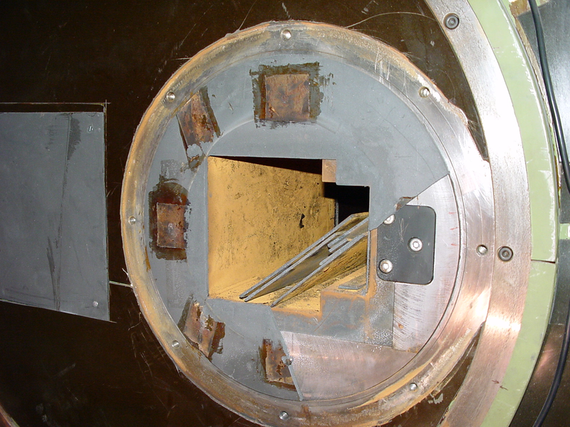



 (jpg - 56 Ki)