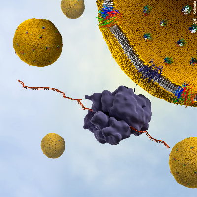
Neutrons reveal hidden secrets of the hepatitis C virus
January 2018
- p7 is a protein essential for the release of the hepatitis C virus, however little data is currently available on the way it interacts with its environment, hindering the development of vaccinations for it
- Researchers have observed the structure of a functional p7 protein within its native environment for the first time using reflected neutrons
- The specific protein insertion mechanism observed will help to outline potential target mechanisms for future drug development.
The hepatitis C virus (HCV) is a blood born virus that causes liver disease and cancer, with more than 300,000 people dying each year and 71 million people living with a chronic infection worldwide. While antiviral medicines are currently used, there is no vaccination currently available and side effects can results in a wrong diagnosis.
In the search to find novel therapies for HCV, researchers have looked to the membrane protein p7, which plays a key role in the release of the virus, for answers. However, there is little data available, and the crystallographic structure of the protein has not yet been resolved.
Recent investigations using neutrons have led to the development of a novel method to study the protein’s integration and structure within a native biological membrane environment. A collaboration between Synthelis SAS, University Grenoble Alpes, and the Institute Laue-Langevin (ILL) enabled researchers to observe the structure of a functional p7 protein complex from HCV for the first time within a physiologically relevant lipid bilayer, at nanoscale resolution.
To do this, the scientists performed neutron reflectometry (NR) on FIGARO, a time of flight reflectometer at the world’s flagship centre for neutron science, ILL in Grenoble, France. Momentum transfer ranges of 0.008> qz> 0.2 Å-1 and minimum reflectivities of R ~ 5x10⁻⁷ were measured using wavelengths λ = 2-20 Å, two angles of incidence and a dqz/qz resolution of 10%.
The Nature Scientific Reports study found that the p7 protein from HCV assembles within the lipid membrane into oligomers that take the shape of a funnel. The conical shape indicates a preferred protein orientation, revealing a specific protein insertion mechanism, and helping to outline potential target mechanisms for future drug development.
As membrane protein dysfunction is also correlated with a wide range of diseases, this advancement in methods to analyse membrane proteins in their native condition, at an atomic scale, also has the potential to help support new therapeutic approaches in other areas, such as for the development of antibodies against HIV.
Erik Watkins, former ILL FIGARO Instrument Scientist, said: “This new approach is a simple and efficient method complementary to other structural and more complex techniques such as NMR and crystallography. This has proved a powerful tool for characterising the protein conformation in its natural environment and one we can look to use for membrane protein discoveries not just in advancements in HCV, but further afield as well.”
Bruno Tillier, Managing Director, Synthelis added: “Neutrons have proved a key resource for this project as we needed to analyse the p7 protein structure in a specific environment. Now we can look to take this deep understanding of the virus not only to devices, like biosensors, but also to study the behaviour of membrane proteins in lipid bilayers to other fields.”
Donald Martin, Head of the research team SyNaBi and professor at University Grenoble Alpes also said: “These new results augur well for our continued development of novel nanostructured systems and devices. The ongoing fruitful collaboration between physicists, biologists and engineers from these institutions in Grenoble provides the important fundamental understanding of physical and biological processes that underlies the development of such nanostructured systems and devices.”
Thomas Soranzo, University Grenoble Alpes and former Synthelis scientist also said: “a major bottleneck in neutron reflectivity analysis of membrane proteins in planar bilayer is the sufficient insertion of polypeptides. This combinatory, new method not only allows significant incorporation of material but also allows specific labelling that could improve membrane protein structure/function studies.”
Instrument: FIGARO - Neutron reflectometer with vertical scattering geometry
Figure 1.
The cell-free preparation of supported bilayers containing p7 and NR and EIS measurements (not to scale). For neutron reflectivity, membranes were formed on quartz and an incident neutron beam was transmitted through the substrate and reflected from Credit: Thomas Soranzo (Synthelis SAS, University Grenoble Alpes), Donald K. Martin (University Grenoble Alpes), Jean-Luc Lenormand (University Grenoble Alpes), and Erik B. Watkins (Los Alamos National Laboratory)
Re.: Coupling neutron reflectivity with cell-free protein synthesis to probe membrane protein structure in supported bilayers, Thomas Soranzo, Donald K. Martin, Jean-Luc Lenormand, & Erik B. Watkins, ScientificReports, 2017
[doi: 10.1038/s41598-017-03472-8]
Notes for editors:
- NR measurements were performed on FIGARO, a time of flight reflectometer at the Institut Laue-Langevin. FIGARO is a high flux, flexible resolution, time-of-flight reflectometer with a vertical scattering plane, optimised for the study of horizontal surfaces such as free liquids. It is possible to strike the interface from above or below the sample in a wide q-range with the instrument. With an incoming beam of wavelengths comprised between 2 Å and 30 Å, and M=4 supermirrors to deflect the beam, it is possible to attain a q-range up to 0.42 Å-1 (reflection up) or 0.27 Å-1 (reflection down) for horizontal samples, and an even broader q-range can be achieved for solid samples which can be tilted using one of the sample goniometers. The sample area includes an active anti-vibration table and free liquid surfaces are aligned automatically using an optical device. The 2D neutron detector allows the acquisition of off-specular scattering and GI-SANS data. You can find out more here.
About ILL – the Institut Laue-Langevin (ILL) is an international research centre based in Grenoble, France. It has led the world in neutron-scattering science and technology for almost 40 years, since experiments began in 1972. ILL operates one of the most intense neutron sources in the world, feeding beams of neutrons to a suite of 40 high-performance instruments that are constantly upgraded. Each year 1,200 researchers from over 40 countries visit ILL to conduct research into condensed matter physics, (green) chemistry, biology, nuclear physics, and materials science. The UK, along with France and Germany is an associate and major funder of the ILL.
About Synthelis
Synthelis is a biotech company based in Grenoble, France. Synthelis’ innovative-patented cell-free technology allows customized expression and characterization of proteins, formulated in proteoliposomes, nanodiscs or in solution with detergent. This makes it particularly suitable for the expression of membrane proteins, which are well known to be challenging to produce in a functional form.
The company offers services in protein production, purification and characterization of difficult-to-express proteins such as ion channel, membrane transporter, GPCR, membrane enzyme, antigen and cytotoxic and soluble proteins. Based on customer-based projects, a protein catalog is also available making cell-free produced proteins readily available.
With on-demand expert services and ever-growing protein catalog, Synthelis is able to support pharmaceutical companies, biotech companies and academic teams in applications such as vaccine development, screening and display technologies, protein delivery, in vitro diagnostics and structural biology.
About Université Grenoble Alpes
The Université Grenoble Alpes (UGA) is the result of a merger in 2016 of Grenoble’s three universities. The UGA, is a comprehensive, global university, offering academic programs and supporting research in all major disciplines: Science, Technology, and Health Sciences; Law, Economics and Business; Humanities and Social Sciences; and Arts, Literature and Languages.
International relations are in the frontline of our global university, which is open to the world and part of a diverse and growing network of partnerships. The UGA’s traditions – of innovation, diversity and excellence – are embodied in our expertise in education, creating a welcoming environment for students, faculty, and staff.
Our academic programs are designed to provide the necessary skills for those who wish to broaden their horizons, whether to meet the challenges of today’s world or to compete in the international job market.
Innovation and excellence also enrich and sustain our world-class research, making the most of an exceptional scientific environment with strong ties to business and industry. Our community of researchers includes experts from all over the world, who work across a large variety of disciplines in the service of knowledge and the spirit of inquiry.


