A deep dive into drug delivery
12 Jun 2026How a novel combination of neutron and light scattering elucidates the molecular structure of therapeutic nanoparticles
-
 Health

Modern medicine increasingly relies on targeted drug delivery - a process during which tiny particles (nanoparticles) transport drugs to specific parts of the body. To ensure these treatments are safe and effective, scientists need to understand exactly how these nanoparticles are built - including their size, shape, and internal structure.
In this study, an international team combined several advanced analysis techniques to investigate drug delivery nanoparticles in unprecedented detail. For the first time, they successfully coupled a particle separation method known as AF4 with small-angle neutron scattering (SANS) during an experiment performed on the ILL’s D11 instrument.
This new approach allowed the researchers not only to measure the nanoparticles’ size and shape, but also to determine how these particles and drug molecules were organised internally. The work opens up new possibilities for using neutron scattering to study increasingly sophisticated drug delivery systems and could help support the development of safer and more effective treatments in the future.
Understanding drug delivery nanoparticles
Modern medicine increasingly relies on targeted drug delivery, a process during which therapeutic molecules are transported directly to specific organs or even certain cell types. To do this, drugs are often packaged inside biocompatible particles (nanoparticles), commonly composed of different fat molecules.
The efficiency of the delivery process is determined by many characteristics of the particles. These include, for example, their internal and external structure as well as the homogeneity of particle sizes within a given batch. International quality standards require that nanoparticle sizes do not vary by more than 30% to be considered safe for application. Close monitoring of particle size distribution is therefore of fundamental importance throughout their manufacturing,
To monitor nanoparticle size, manufacturers commonly use a technique called asymmetric-flow field-flow fractionation (AF4). In brief, it involves separating particles in solution in such a way that smaller particles move faster than larger ones. AF4 is usually coupled with methods such as ultraviolet light absorbance or light scattering in order to measure the amount of particles in each size group.
A powerful new combination of techniques
To understand nanoparticles even better, researchers also need information about their shape and internal structure. This requires combining AF4 with techniques specifically designed to probe how particles are organised at the nanoscale.
In previous studies, AF4 has been coupled to small-angle X-ray scattering (SAXS) to investigate tiny magnetic particles. However, it had never before been combined with neutron-based techniques such as small-angle neutron scattering (SANS).
This challenge has now been overcome by an international team including scientists from the Leibniz Institute for Polymer Research (Dresden), Stellenbosch University, Max IV and the Department of Process and Life Science Engineering (Lund) and the ILL, who successfully carried out the world’s first AF4-SANS experiment on the ILL’s D11 instrument.
Using nanoparticles designed for drug delivery, the team analysed them with an AF4 setup coupled simultaneously to Multi-Angle Light scattering and, for the first time, SANS. Using this powerful combination, the researchers were able to determine not only the dimensions of the particles, but also to test the homogeneity of their internal structure and the potential location of drug molecules with great precision. This combined approach gave the researchers a far more detailed understanding of how the nanoparticles are built.
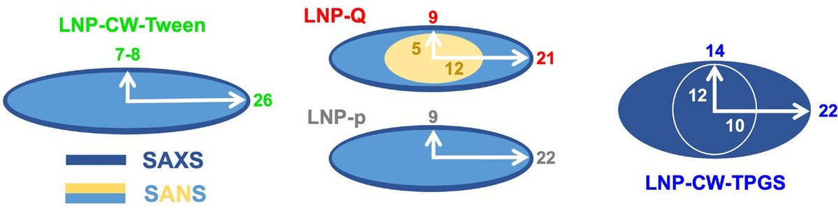
Figure 1: This schematic shows how SAXS and SANS revealed complementary details about different drug delivery nanoparticles. SAXS helped determine the overall particle shape and dimensions, while SANS provided additional contrast to probe the internal structure. The numbers indicate dimensions in nanometres. The different structures reflect differences in nanoparticle composition: Tween and TPGS are surfactants used to stabilise the particles, and the study showed that changing these surfactants can alter particle shape, size distribution, and internal organisation. Credit: Small Methods (2026)
In the Polymer Separation Group at Leibniz Institute of Polymer Research Dresden, we have pioneered the coupling of advanced field-flow fractionation techniques with powerful scattering methods, including AF4-SANS and thermal FFF-SAXS. These multidetection approaches allow us to extract complementary, orthogonal information from very small amounts of sample, which is particularly valuable in biomedical research. Looking ahead, we believe that such integrated analytical strategies will be decisive for understanding complex polymer systems and for guiding the design of next-generation polymer-based biomedical applications.
"Prof. Dr. Albena Lederer"
Overcoming experimental challenges
The team also introduced an important improvement to the standard AF4 setup. During AF4 experiments, particles become diluted as they move through the system, which weakens the measurement signal and can lead to very long experiment times.
To overcome this problem, the researchers implemented a method to compensate for this dilution effect, allowing them to obtain reliable measurements much more efficiently. This included focusing the detection of the nanoparticle signal exclusively on the nanoparticle-rich solution, while directing the solvent signal away from the detectors and allowing for signal detection with good statistics.
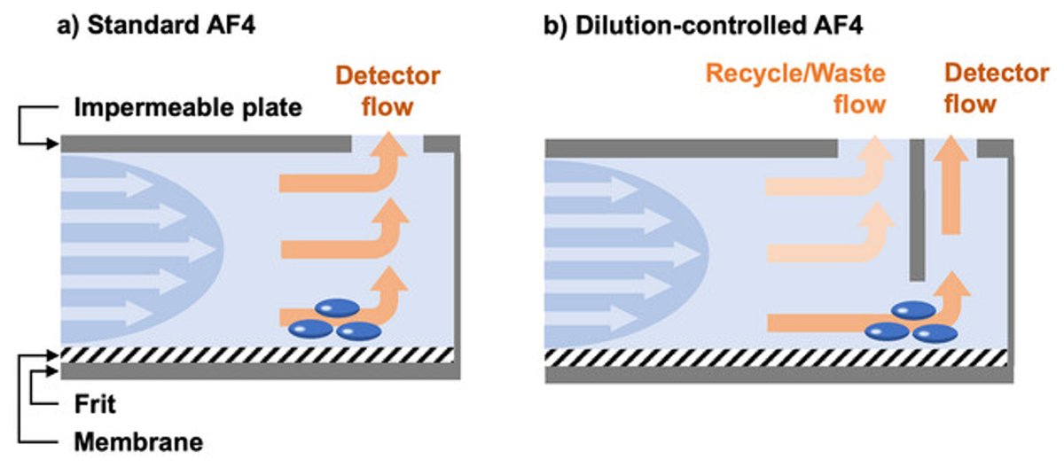
Figure 2: In standard AF4 setups (left panel) both nanoparticles and solvent reach the detector simultaneously. This weakens the signal originating from the nanoparticles, thus increasing measurement time. In the dilution-controlled setup, pioneered in the publication described here, much of the solvent-rich flow is diverted to waste, resulting in a stronger nanoparticle signal (right panel). Credit: Small Methods (2026)
A new role for neutrons in drug delivery research
The coupling of SANS to AF4 is an extremely important step in the development of nanoparticle characterisation platforms. One major advantage of neutron scattering is its sensitivity to hydrogen and deuterium. By selectively replacing hydrogen atoms with deuterium, researchers can make specific parts of a nanoparticle stand out, revealing structural details that are otherwise difficult to observe with other techniques.
As targeted treatments continue to develop, precise characterisation of drug delivery nanoparticles is becoming increasingly important. The experimental framework established in the study described here is an important contribution to tackling this challenge, and opens new possibilities for the use of neutron scattering in biomedical research.
Reference:
Bittrich, E., Boye, S., Van Niekerk, Z., Stanvliet, Z., Porfetye, A., Herranz‐Trillo, F., Bolinsson, H., Gaydarova, S., Tzachev, C., Martel, A. and Nilsson, L., 2026. Structural Profiling of Lipid Nanoparticles at Sub‐10 nm Resolution via AF4 Coupled Online to SAXS and SANS. Small Methods (2026) https://doi.org/10.1002/smtd.70639
ILL Instrument: D11
ILL Contact Persons: Anne Martel, Ralf Schweins
Institutions involved in the research: Leibniz-Institut für Polymerforschung Dresden, Stellenbosch University, Wyatt Technology Europe, MAX IV Laboratory, Lund University, Sofia University St. Kliment Ohridski