

How do wounds heal? Neutrons enable a discovery that reshapes our understanding of the process
20 Aug 2026Scientists have redefined how the key blood-clotting protein fibrinogen behaves when it comes in contact with air, overturning two decades of scientific consensus on wound healing. Neutron reflectometry measurements at the ILL played a key role.
-
 Health

On the processes of science
Breakthrough science results do not appear out of thin air. Besides invariably starting with the ability to ask the right questions, they usually involve years of step-by-step discoveries and encompass collaborations between scientists who combine their skills and knowledge. Last but not least, they are often driven by technology developments – new state-of-the-art tools and methods that come into play and shed some light on what was obscure or invisible before. This study is a paradigmatic example of this process.
Do we really know how it works?
Fibrinogen is a large blood-clotting protein essential for the body's natural process that stops bleeding when a blood vessel is damaged, as well as sealing open wounds when blood comes in contact with air. But how well do we know the process of scab formation on the surface of blood in open wounds?
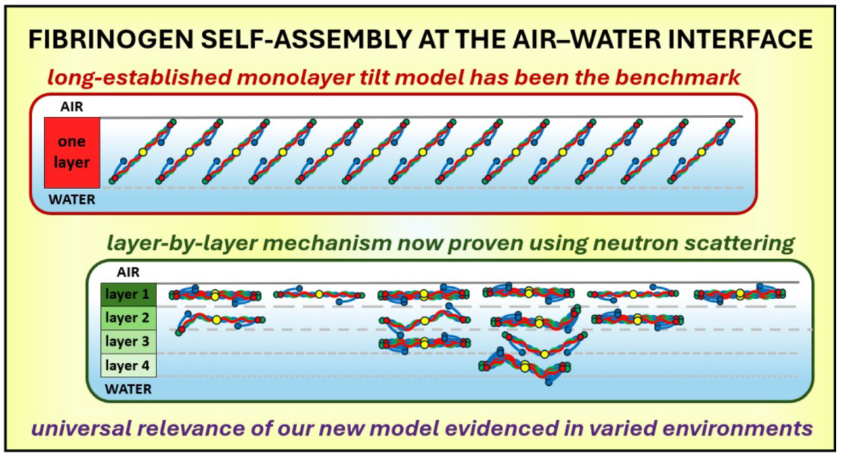
For more than two decades, the field has relied on a widely accepted ‘single tilting layer’ model for how fibrinogen molecules organise when they come in contact with air. The long fibrinogen molecules were thought to organise in a monolayer, lying flat on the surface at first, and then gradually tilting upright as more of them arrived. In other words, at low concentrations the long axis of the fibrinogen molecules would be parallel to the surface, forming a single thin layer (about 5 nm), while with increasing concentrations the molecules would tilt until the long axis was perpendicular to the interface, forming a single thicker layer (46 nm when saturated).
The model was based mainly on optical measurements using ellipsometry, a technique that measures the change in light polarisation after reflection from a sample, and which can indirectly infer layer thickness and other characteristics by comparison with an optical model. The ‘single tilting layer’ model persisted as a benchmark for decades, until scientists decided to ask the question again: do we really know how it works?
State-of-the-art techniques to test an old model
The approach of the authors of the new study was to revisit the long-established model in the light of new knowledge (about how large protein molecules interact in solution) and state-of-the-art, surface-sensitive experimental techniques – namely neutron reflectometry.
Neutron reflectometry involves shining a collimated beam of neutrons onto a flat surface and detecting the reflected neutrons. The technique is non-invasive, and reflectivity profiles provide detailed information about the composition and structure at fluid interfaces. Neutron reflectometry measurements were conducted on the time-of-flight neutron reflectometer FIGARO at the Institut Laue-Langevin (ILL).
"It's never easy challenging an established model of how molecules behave in nature. But through using the advanced neutron reflectometry technique on the FIGARO instrument at the ILL research facility, we could see the structure of these protein surfaces in more detail than scientists had seen before,” explains principal investigator Dr Richard Campbell, who worked at ILL when the initial experiments were conducted, and is now a Senior Lecturer at the University of Manchester.
A fundamentally different mechanism
The study, “Redefining Fibrinogen Self-Assembly at the Air–Water Interface: An Intriguing Story with Multiple Layers”, published today in the Journal of the American Chemical Society is the culmination of over a decade of international collaboration between scientists in France, the UK, Spain and Chile. It shows that the benchmark model missed the fact that fibrinogen stays lying flat and builds multiple layers that stack like sheets of paper. These layers grow thicker and more complete as more molecules arrive. This new information helps to explain how the long protein molecules line up on the surface of blood where fibres called fibrin form the basis of the scab, a solid film formed during evaporation of fluid at the blood surface, that seals a wound.
The team demonstrated that this behaviour holds true across multiple ranges of concentration and in very different solution conditions, showing that the mechanism is a universal feature of fibrinogen when it comes in contact with air. Interestingly, a single layer of perpendicularly oriented molecules would promote more intense and cohesive interactions, and consequently more elasticity, which is not confirmed by dedicated measurements. The new model is far more compatible with what is observed in practice.
Far-reaching implications
“Having better understanding of the structure of fibrinogen at liquid surfaces can only help in the search for new and better treatments for patients whose wound refuse to heal,” says Campbell.
Indeed, the discovery has far-reaching implications for health science. Elucidating the mechanism of scab formation on the surface of blood in open wounds is of particular importance to patients who take anticoagulant medication, or have autoimmune disorders that prevent wounds from healing. The findings could also help explain aspects of how a lung can collapse in patients who suffer from acute respiratory distress syndrome, as fibrinogen disrupts the function of the lipid layer that keeps the airways open during breathing. They may also inform the development of biosensors which allow people with diabetes to monitor how the glucose content in their blood is performing, as fibrinogen disrupts their function as well.
Reference:
G. J. Coope, M. J. Lawrence, P. Gutfreund, N. Hassan, J. M. Ruso and R. A. Campbell “Redefining Fibrinogen Self-Assembly at the Air–Water Interface: An Intriguing Story with Multiple Layers.” Journal of the American Chemical Society, 2026 https://doi.org/10.1021/jacs.6c11928
ILL Instruments: ⚙️FIGARO
Contact Persons: Richard Campbell (U. Manchester) and Philipp Gutfreund (ILL)
Institutions involved in the research: University of Manchester, Universidad de Santiago de Compostela, Lund University, Universidad Tecnológica Metropolitana and Universidad de Valparaíso
This work was featured in the Financial Times on Friday, 21 August 2026.
It also appeared here: https://phys.org/news/2026-08-fibrinogen-discovery-reshapes-wounds.html, https://streamlinefeed.co.ke/news/groundbreaking-neutron-scanning-discovery-upends-50-year-medical-consensus-on-wound-healing