
Page 39 - ILL Annual Report 2019
P. 39
SCIENTIFIC HIGHLIGHTS
36-37
250 K
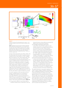
Figure 2
Schematic illustration of the probed length and time ranges, i.e. the momentum-energy (Q-E) space of the five different instruments used in this study.
Inset) The dynamical structure function map, S(Q,E) of deuterated RR-P3HT (d-RR-P3HT) measured at 250 K on the IN6 spectrometer. The elastic signal, S(Q), of d-RR-P3HT measured at 296 K using the D16 diffractometer is shown on the left-hand side. The Q-averaged QENS signal, S(E), of d-RR-P3HT from IN6 is depicted on the bottom left-hand side, and the inelastic (INS) signal, S(E), of d-RR-P3HT measured at 10 K on the IN1-Lagrange spectrometer, is depicted on the bottom right-hand side.
processes and tune the probe using contrast variation. This not only provides information about co-operative motions and self-motions but also enables us to hide or highlight part of the molecules. Third, neutron spectroscopy is one of the few techniques that allows the probing of both structure (Q-range or space) and dynamics (energy or time range) on the femtosecond to nanosecond time scale. Most exciton/charge transfer processes in conjugated polymers occur on the femtosecond to picosecond time scale. Charge transport, despite occurring by means
of a hopping process and being characterised by a longer time scale of hundreds of picoseconds to tens of nanoseconds, results from charge transfer involving local energy sites. Thus, neutron spectroscopy allows us to cover the time scale relevant to optical and electronic processes.
We used a combination of five instruments at the ILL in synergy—the diffractometer D16, the cold-neutron time-of-flight spectrometer IN6, the backscattering spectrometer IN16B, the spin-echo spectrometer IN11 and the hot-neutron spectrometer IN1-Lagrange—to obtain a full map of the structural dynamics of a conjugated polymer model system poly(3-hexylthiophene) (figure 2):
(i) We used neutron diffraction to primarily probe the crystalline/ordered structure of the polymer. Local and
vibrational dynamics were probed using synergistically
different neutron spectroscopic techniques.
(ii) Time-of-flight, backscattering and spin-echo
measurements were performed to probe local and relaxation dynamics by covering both the picosecond and nanosecond time scales. These techniques allowed us to probe the structure (Q-range) and the dynamics (energy range) simultaneously and thus to gain deeper insight into the relationship between the two. Further insight into the dynamics was gained through deuteration and molecular dynamics (MD) simulations. MD enabled us to calculate both coherent and incoherent intermediate scattering functions.
(iii) Hot-neutron vibrational spectroscopy was carried out to map out the full vibrational spectrum, including both the lattice (external) modes and the molecular (internal) degrees of freedom. Vibrational spectroscopy is not only sensitive to ordered phase but also to disordered phase. Thus, by combining neutron vibrational spectroscopy with MD, molecular and periodic density functional theory calculations, we bridged the gap between the molecular level and the solid-state scale.
This study [2] allowed us to put specific emphasis on the impact of disorder, degrees of freedom and packing, and on investigating the adequacy of the theory needed to describe and capture the essential structural and dynamical features of the conjugated polymers studied. Different length scales and time scales were appropriately covered, allowing us to describe the various phases and energy landscapes of the studied materials adequately. This is of the utmost importance, since tailoring materials for efficient energy applications depends on a better understanding of the underlying microstructural phases and the associated dynamical behaviours.
www.ill.eu
