
Page 17 - ILLS Annual Report 2018
P. 17
SCIENTIFIC HIGHLIGHTS
14-15
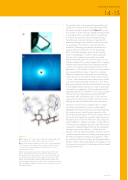
Figure 2
a) The galectin-3C crystal used to collect data for the complex with lactose at LADI. The volume of this crystal was 1.8 mm3.
b) Part of the diffraction pattern from this crystal, which extended to 1.7 Å resolution. The increase in the crystal size from 0.4 mm3 to 1.8 mm3 reduced data collection time by a factor of 3.5 and increased the resolution.
c) The nuclear density map for the complex of galectin-3C with lactose. The protonation states of all amino acid side chains in
the binding site are clearly defined, as are three critical hydrogen bonds from hydroxyl groups on the upper face of lactose to the protein. The high degree of tailoring of the protein surface to the exact hydrogen-bonding pattern of the natural disaccharide confirms that drug design strategies have been wise to include this core in all developed inhibitors.
The relatively polar, surface-exposed ligand binding site of galectin-3 contains several residues that interact with the ligands through hydrogen bonds (figure 1), as well as a number of water molecules. Despite it being possible to consistently obtain very high-resolution X-ray data (up
to 0.86 Å), the geometries of key hydrogen bonds in the binding site remained ambiguous. Experimentally determined hydrogen atom positions could also provide an unambiguous foundation for molecular dynamics simulations, free-energy perturbation calculations and quantum chemical calculations in future drug design efforts. To find the hydrogen atoms, we thus turned to neutron crystallography. In order to obtain good data from our initial, modestly-sized crystals, we worked
with perdeuterated galectin-3C from the outset. Our first diffraction patterns from crystals of galectin-3C in complex with the natural ligand lactose were obtained at LADI-III
in 2012, from crystals measuring about 0.35 mm3 that diffracted to 1.9 Å resolution. However, the data collection times were prohibitively long: 16 days for a complete dataset. By optimising crystal growth over the following couple of years we were able to obtain crystals of up to 1.8 mm3, which reduced the data collection time at LADI to six days and increased the resolution to 1.7 Å. These data were combined with data from the MaNDi instrument at Oak Ridge National Laboratory in order to obtain data of exceptional completeness. We also obtained data for ligand-free galectin-3C at LADI-III in 2016, and a complex with the non-natural ligand glycerol from the BIODIFF instrument at MLZ in Garching in 2015. Thus, the final analysis built on data from a number of neutron sources, although the data from LADI-III were of key importance.
The neutron crystal structures of galectin-3C in complex with lactose and glycerol, as well as without ligand, highlighted the exquisite fine-tuning of hydrogen bonding from protein atoms to match the exact geometry of the natural disaccharide ligand. All the polar oxygen atoms on the ‘inside’ of the ligand are involved in highly directional hydrogen bonds. A common method for discovering new protein inhibitors is ‘fragment screening’, in which libraries of small molecular fragments (typically under 300 Da) are screened for weakly-binding hits that are then developed into larger, more potent inhibitors. It is likely that such libraries, which are often dominated by aromatic moieties, would be especially effective in finding new drugs against the shallow, hydrophilic binding sites of galectins, which are exquisitely tailored to recognise the hydrogen-bonding patterns on their carbohydrate ligands. It thus appears wise to continue the current trend of developing inhibitors based on disaccharide cores.
www.ill.eu
