
Page 16 - ILLS Annual Report 2018
P. 16
BIOLOGY AND HEALTH
Derek Logan. British and Swedish Department of Biochemistry and Structural Biology, Lund University, Sweden
‘We’ve been using neutrons at the ILL and other sources for six years now, after two-and-a-half decades of purely X-ray crystallography. This has opened up a whole new world for me.’
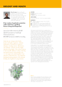
First study of galectin proteins with neutrons, guiding
future drug development
Quasi-Laue diffractometer LADI-III
MaNDi instrument at Oak Ridge National Laboratory
BIODIFF instrument at MLZ in Garching
Galectins are a large protein family
found in all higher organisms. Galectin dysfunction is involved in a variety of diseases, including cancer, inflammation and diabetes. Galectins are thus highly attractive drug targets. One common feature of all successful inhibitors to date is that they are based around a disaccharide core,
i.e. two sugar units joined together. In an attempt to understand why, we probed the hydrogen-bonding patterns in the substrate binding site of galectin-3 using neutrons.
AUTHORS
D.T. Logan (Lund University, Sweden)
M. Blakeley (ILL)
E. Oksanen (ESS-ERIC, Lund University, Sweden)
ARTICLE FROM
J. Med. Chem. (2018)—doi: 10.1021/acs.jmedchem.8b00081
REFERENCES
[1] L. Johannes, R. Jacob and H. Leffler, J. Cell. Sci. 9 (2018) 10.1242/jcs.208884
[2] G.A. Rabinovich et al., J. Scand. Immunol. 66 (2007) 143 [3] G. Pugliese et al., Clin. Chem. Lab. Med. 52 (2014) 1413 [4] D. Laaf, P. Bojarová, L. Elling and V. Křen, Trends Biotechnol
(2018) Nov 6. pii: S0167-7799(18)30264-6—doi:10.1016/j. tibtech.2018.10.00
The ubiquitous protein family known as galectins has 15 members in mammals, and is defined by the affinity that all its members have for glycans containing β-D-galactoside groups [1]. One of the most studied members is human galectin-3, found in both the nucleus and the cytoplasm of cells, and also secreted to the outside of cells where it interacts with β-galactoside-containing glycoproteins and glycolipids,
e.g. in glycosylated proteins. Galectin-3 consists of two domains. The N-terminal one is involved in higher order oligomerisation, a characteristic unique among galectins [1], through an as yet unknown mechanism. This is followed
by a C-terminal carbohydrate-recognition domain.
Human galectin-3 is a current pharmaceutical target as it is involved in various major diseases, such as inflammation, cancer proliferation and metastasis, and diabetes [2, 3]. The galectin-3 inhibitor Td139 is about to enter phase II/III clinical trials for the treatment of idiopathic pulmonary fibrosis. We and others have determined many high-resolution X-ray crystal structures of the C-terminal domain of galectin-3 (galectin-3C) in complex with synthetic ligands, as part of the structure- based design of new inhibitors [4]. The most successful of these are invariably based on the natural substrate lactose,
or the very similar dithiogalactoside. Instead of modifications to the disaccharide core, affinity has been enhanced through the exploration of additional adjacent binding pockets.
We wanted to understand why the disaccharide core is so important in terms of hydrogen-bond directionality.
Figure 1
Binding of the drug candidate Td139 to the carbohydrate recognition surface of galectin-3 (from PDB entry 5H9P). The Td139 molecule
is shown in stick representation, as are important residues in the shallow binding groove involved in interactions with the ligand. The disaccharide core on which all the most potent galectin-3 inhibitors to date are based is indicated by an oval.
ANNUAL REPORT 2018
