IN1-Lagrange
Large Area GRaphite ANalyser for Genuine Excitations
Reactor hall, hot beam H 8 |
|---|
beam size: 20 cm height x 5 cm width |
Monochromators | |||
|---|---|---|---|
size: 20 cm height x 18 cm width | |||
crystal | spacing d/Å | Ei range (meV) | ΔEi (meV) |
Cu(220) | 1.278 | [26-500] | 2-3% Ei |
Cu(331) | 0.829 | [67-500] | 1.5-2% Ei |
Si(311) | [16.5-60] | 0.8 | |
| Si(111) | [4.5-20] | 0.8 | |
Collimation |
|---|
Boron coated collimators |
α2, α3, α4 = 20', 30', 40', 60' |
Sample |
|---|
beam size at sample position 3x3 cm2 |
Analysers |
|---|
Focusing reflecting surface of 1 m2 built around the vertical sample-detector axes from PG002 crystals. Ef= 4.5 meV |
3He detector |
|---|
Instrument description
N1 works in a time-sharing mode. This means that the same monochromator is also used by IN1 and by the liquids diffractometer D4. Changing over between the three different instruments can be done without difficulty in about two hours.
Either the three-axis spectrometer (TAS) and IN1 Lagrange. Spectrometer can be connected to the instrument monochromator unit. The secondary spectrometers are mounted on "Tanzboden" modules with the possibility of varying the distance between the different modules. The interchange of these two secondary spectrometers does not take long time - about an hour. The two spectrometers of IN1 operate on the basis of time sharing of the common monochromator with the liquids diffractometer D4.
The monochromator unit carries three different double focussing monochromators built from copper single crystals (available reflecting planes Cu(220) and Cu(331), Si(311) and Si(11)). The exchange of the monochromator planes is controlled by the instrument computer. The radius of curvature can be automatically adjusted as function of reflected energy in order to maintain maximal flux at the sample position in the course of energy scans.
The scattering angles on the monochromator cover a range of 10°<2M <90° allowing for scanning neutron energies from ~ 4-5 meV to more than 1 eV (see table of characteristics).
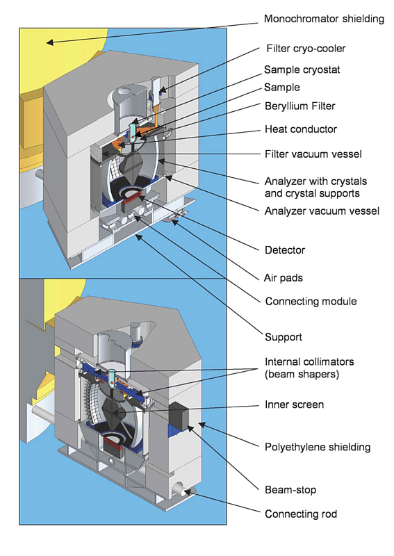
The IN1-LAGRANGE (LArge GRaphite ANalyser for Genuine Excitations) secondary spectrometer setup (Fig. 1) is based on the space focussing of neutrons scattered by the sample in a very large solid angle, which are all then recorded with a relatively small single counter (a He3 gas detector). The focussing reflecting surface of ~1 m2 is built around the vertical sample-detector axis from pyrolytic graphite (PG) crystals set to reflect neutrons with the fixed average energy of 4.5 meV. Altogether there are 612 crystal holders mounted on the solid mechanical structure so that the individual flat crystals are set tangentially to the calculated ellipsoid of rotation. The crystal sizes, their mosaic spread and the height of the part covered by PG crystals are optimised in a way such that all three corresponding contributions to the energy resolution are equal. It is important that the requested energy resolution of the instrument is achieved with a relatively low grade of the pyrolytic graphite. The appropriately shaped beryllium filter is installed immediately after the sample in order to remove higher-order harmonics in the analyser reflections. The filter transmission below the cut-off is enhanced by cooling it to the base temperature (20 K) of a powerful closed-cycle helium refrigerator. The resulting accepted solid angle (~0.8π ≈ 2.5 Steradian) is among the highest apertures open for scattered neutrons in the most ambitious instrument projects at present. At the same time it is realised within the highly compact characteristic spectrometer volume, about 1 m3 for the analyser + filter pumped volumes). The carefully designed screen of boron-containing absorber is installed on the sample-detector axis in order to suppress the intense elastic scattering from the sample. Further reduction of the instrument background, contaminated with the high neutron energy components, is achieved through massive polyethylene shielding built around the whole analyser.
The IN1-TAS spectrometer: the scattering angles at the sample and the analyser can be changed in the intervals -115°<2θS<115° and -120°<2θA<120°. Three different analysers (PG(002), Cu(200), Cu(220)) can be installed in order to optimise intensity and resolution for a given experiment.
Various resonance absorption filters (e.g. Er, Sm, Hf ...) can be used to suppress higher order contaminations from the incident beam or in the scattered beam. An oriented Pyrolytic Graphite filter is designed for experiments eventually demanding thermal neutron energy range.
 (jpg - 246 Ki)
 (jpg - 159 Ki)