
Page 58 - ILL Annual Report 2019
P. 58
BIOLOGY AND HEALTH
Svetlana Antonyuk. British Department of Biochemistry, University of Liverpool, UK.
‘We’ve being using neutrons since 2008,
in addition to our synchrotron and XFEL experiments. Neutrons have allowed us to look at our enzyme from a different perspective.’
Neutron structure of a denitrifying enzyme provides new insights into the catalytic mechanism
Quasi-Laue diffractometer LADI-III
Copper nitrite reductases are a family
of metalloenzymes that convert nitrite
to nitric oxide in the denitrification pathway. As such, they are of central importance in nitrogen-based energy metabolism. To gain further insight into the enzyme mechanism we used neutron crystallography. This revealed unexpected protonation states for key residues and important details concerning hydration.
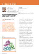
Figure 1
Ribbon diagram of the AcNiR trimer. The copper ions are shown as dark blue spheres.
AUTHORS
S.V. Antonyuk (University of Liverpool, UK)
M.P. Blakeley, M. Moulin and M. Haertlein (ILL)
ARTICLE FROM
IUCrJ. (2019)—doi: 10.1107/S2052252519008285
REFERENCES
[1] S.V. Antonyuk et al., Proc. Natl. Acad. Sci. USA 102 (2005) 12041 [2] M.P. Blakeley, S.V. Antonyuk and S.S. Hasnain, IUCrJ. 2 (2015) 464
Denitrification is the process by which some micro-organisms couple respiratory ATP synthesis with the reduction of nitrite (NO2−) to dinitrogen (N2) via the gaseous product nitric oxide (NO). Denitrification forms an important step in the global nitrogen cycle. It has agronomic, environmental and medical impacts. The individual reactions of denitrification are catalysed by distinct reductases that can contain a variety of metal or heme centres.
Copper-containing nitrite reductases (CuNiRs) are involved in the first step of the denitrification pathway, catalysing the reduction of nitrite to nitric oxide in a one- electron two-protons reaction. The biological assembly
is a trimer, with each monomer containing two distinct types of copper (Cu) centre (figure 1). The type 1
Cu ion (T1Cu) site is responsible for transferring an electron to the type 2 Cu (T2Cu) site, where catalysis occurs. The T2Cu is co-ordinated by three histidine
(His) residues, plus a ligated water that is displaced when nitrite binds to the oxidized T2Cu. Two additional residues located close to the T2Cu site are known to be essential for enzymatic activity: an aspartic acid (Aspcat) and another histidine (Hiscat). Aspcat is hydrogen-bonded to the T2Cu ligated water and also to Hiscat via a bridging water.
A number of atomic resolution structures of CuNiRs from the soil bacterium Achromobacter cycloclastes (AcNiR) were determined using X-rays, including oxidized and reduced forms as well as nitrite-bound complexes [1, 2]. However, despite a large proportion of hydrogen (H) atoms being visible, many of the key H atoms of the catalytic T2Cu site were not. Moreover, like all metalloenzymes, AcNiR is very susceptible to radiation damage from X-rays, imposing a serious limitation on unravelling details of the resting state of AcNiR at the atomic level. For this reason, in parallel to our synchrotron and XFEL studies we chose to use neutrons, since they offer a way to determine damage-free structures of enzymes at room temperature and can provide details of protonation and hydration, necessary for determining catalytic pathways.
ANNUAL REPORT 2019
