
Page 51 - ILL Annual Report 2019
P. 51
SCIENTIFIC HIGHLIGHTS
48-49
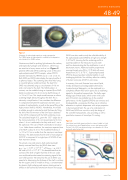
Figure 1
Schematic of sophorolipid-coated iron oxide nanoparticles.
The COOH group of sophorolipids is modified by nitrodopamine, which binds to the SPION surface.
Neutrons are ideal for probing light elements like carbon, and especially hydrogen and deuterium, while X-rays
are sensitive to heavy atoms such as iron. Figure 2 presents SAXS and SANS scattering curves of the same sophorolipid-coated SPION sample, whose SPION diameter estimated by HR-TEM was d = 4.6 ± 0.3 nm. Both SAXS and SANS data display the typical profile of a spherical object. The scattering data were fitted using a core-shell sphere model (red lines). The best fit for
SAXS was obtained using a core diameter of 4.6 nm and a null value for the shell. The SANS pattern, in contrast, can be modelled using an imposed (from SAXS data) core diameter of 4.6 nm and a shell thickness of
t = 2.6 ± 0.2 nm. This simple model assumes a uniform scattering length density throughout the shell. This is obviously a simplification if one considers the difference
in composition between the sophorose and oleic acid moieties of sophorolipids, as well as the space-filling of the high-curvature shell by NDA-SL. Nonetheless, this model
is reliable enough to estimate the shell thickness by fitting the shift of the first minimum of the form factor observed
in the SANS compared with the SAXS scattering curves. The calculated length of SL, given the 120 ° angle due to the mono-unsaturation of SL, can be estimated at 2.6 nm, where 1.6 nm is attributed to the fatty acid and 1 nm to the sophorose. The size of NDA is estimated to be about 1 nm. Under these conditions, the maximum expected size of SL NDA is about 3.6 nm. The modelled thickness of
t = 2.6 ± 0.2 nm is smaller than the maximum monolayer thickness. This demonstrates that the shell of monodisperse SL-IONP is composed of a single layer of SL-NDA of less than maximum density.
SANS was also used to study the colloidal stability of the sophorolipid-coated SPIONs at high ionic strength (1 M NaCl), showing that the scattering profile is superimposable on that measured in pure water
and thus demonstrating that the stabilisation is not of electrostatic nature. Additional stability experiments were performed using DLS in 10 % fetal calf serum heated from 20 °C to 70 °C. Sophorolipid-coated SPIONs showed excellent colloidal stability in such challenging biofluids. Nor did they affect the viability of human monocytes (U937) in cell culture.
In summary, this work illustrates how natural lipids, initially developed as biodegradable alternatives
to petrochemical detergents, can be employed in a completely different field and in particular as stabilising agents for biomedical nanoparticles. The bulky sugar headgroups of these lipids, which orient in the outer
part of the shell, are probably key to the excellent performance. As the SL-grafted SPIONs are non-toxic and biodegradable, we propose that they are an attractive alternative to synthetic dispersants with unique properties in the biomedical field. The use of a combination of SANS and SAXS was crucial to verify the design of
the core-shell nanoparticles, also providing us with a quantitative measure of monolayer SL coating.
Figure 2
SANS (blue) and SAXS (green) profiles for 4.6 nm SL-IONP in D2O. The red lines show the fit obtained from a core−shell sphere model. The best fit for SAXS was obtained using a core diameter of 4.8 nm and a null value for the shell.
The solvent−core and solvent−shell scattering length
density contrasts fitted from the SANS data were
ρsolv – ρc = –0.57 × 10⁻⁶ Å–2, while ρsolv – ρc was in the order of 3.0 × 10⁻⁶ Å–2. This demonstrates that the contrast of the shell is higher than that of the iron oxide core and therefore dominates the scattering signal of the core−shell particle. We stress that the use of SANS was essential in determining the thickness of the shell in solution: the thin organic shell is invisible to X-rays, is overestimated by dynamic light scattering and requires staining prone to artifacts to be resolved by transmission electron microscopy.
www.ill.eu
