
Page 21 - ILLS Annual Report 2018
P. 21
SCIENTIFIC HIGHLIGHTS
18-19
a) [111]
Elastic Recovery Force
b)
3D Simulations (ECT)
Experimental reconstructions
Stray field lines
Magnetic Force
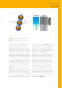
Figure 2
a) Schematic representation of the forces involved in the construction of the chain.
b) Experimental reconstruction from ECT images and 3D simulation of the chain.
The magnetosome chain is a natural paradigm of 1D magnetic nanostructure. Due to the large magnetic anisotropy, such arrangements show potential for biomedical applications [2] and actuation devices such
as nanorobots [3]. Rather than displaying straight lines, magnetosome chains are slightly bent, as demonstrated by electron cryotomography (ECT). This complex structure must have an effect on the magnetic behaviour of the chain. The present study was devoted to shedding light on the underlying mechanisms that determine the arrangement of the magnetosomes and, consequently, the geometry of the chain. With this aim we explored, on a bacterial colloid, the orientation of the magnetic moments using the small- angle neutron scattering instrument D33 with longitudinal neutron-spin analysis (POLARIS). The major advantage of using POLARIS lies in the proper separation of coherent nuclear and magnetic scattering by measuring the two non-spin-flipped (nsf) intensities and two spin-flipped (sf) cross-sections.
Figure 1a presents the purely nuclear scattering intensities Inuc(q) determined by integration of the nsf channels in 10 ° sectors along the applied field direction for two different field strengths of μ0H = 2 mT and 1 T, respectively. The indirect Fourier transform of Inuc(q) results in the pair- distance distribution function P(r) displayed in figure 1b. The nuclear intensities Inuc(q) and P(r) for the two fields are identical, showing that at 2 mT the bacteria are already
fully aligned in the field direction (see figure 1c). On the other hand, the cross-term Icross(q) obtained by integration of I −−(q)−I ++(q) in 10 ° sectors around Θ = 90 ° (q⊥H) yields information on the polarisation-dependent nuclear- magnetic interference scattering [4]. The cross-terms Icross(q) detected at 2 mT and 1 T (figure 1a) and the extracted distribution functions (figure 1b) display the same functional form. However, the absolute values of P(r) at 2 mT over the whole r-range are reduced by a factor of 0.83 compared with a saturating field of 1 T. In combination with isothermal magnetisation measurement, these results suggest two individual steps in the magnetisation process: chain alignment, followed by coherent rotation of the magnetic moments within a magnetosome into the field direction. At 2 mT, the nanoparticle magnetisation deviates by 20 ° from the chain axis, which coincides with the crystallographic easy axis of magnetite along [111].
The direction of the net magnetic moment, which remains along the chain axis, is not affected. However, the tilt turns out to be the key to understanding the arrangement of magnetosomes in helical-shaped chains. The chain shape can be reproduced by considering an interplay between magnetic dipole-dipole interactions, ruled by the orientation of the magnetosome magnetic moment, and a lipid-/protein-based elastic recovery force caused by the connection of the magnetosome with cytoskeletal filaments that traverse the cell (see figure 2).
www.ill.eu
